Athena Review Vol. 5, no. 1
Records of Life: Fossils as Original Sources
2. Early views of fossils
Much of the history of life has been recorded in fossils- which are themselves a kind of happy coincidence, literally the accidental casts or reliefs of ancient plants and animals. Silt, clay, fine-grained sand, and dissolved minerals such as silica or iron often gradually replace the actual once-living parts of buried organisms in casts formed by surrounding dirt. In time the dirt becomes sedimentary rock, containing the fossils. These occur either as positive or negative casts- the former, such as a shark’s tooth, a dinosaur leg bone, or a trilobite made out of rock; the latter, such as the cast of a mollusk shell, leaving in the rock a negative relief or depression (fossa in Latin, hence the term “fossil”). As in the case of microbial life during the Precambrian period, in the smoothest rock such as chert, even the tiniest of microorganisms may be preserved as fossils, and can be recorded by modern microscopic imagery.
Since ancient times, people have noticed fossils in all kinds of sedimentary rocks during their daily lives. Fossils easily visible to the naked eye are common in the sedimentary rocks used for building stones, including limestone, sandstone, and shale, the earliest dating back to the Cambrian and Ordovician periods (542-450 million years ago). Another common source of fossils is coal, showing often well-preserved plant remains from the Carboniferous era (about 360-290 million years ago).
The most consistent awareness of fossils as ancient life forms (as opposed to mere rock inclusions, which coincidentally resembled forms of life) seems to have come when the fossils of familiar organisms are seen far from their normal setting. Sea shells, for example, are frequently observed in rock strata far from the sea. If sea shells are seen in rocks layers at a setting many miles inland, one possible conclusion is that the environment must have changed after they were deposited . Such conclusions, we know, were independently reached and reported by the ancient Greek philospher Xenophanes, the Song Dynasty Chinese astronomer Shen Kuo, and the Italian Renaissance artist Leonardo da Vinci, among others. All three of these persons were independent thinkers who were little constrained by standard explanations of their times, which in da Vinci’s day included the idea that the Biblical Flood had moved such marine shells around a few thousand years ago..
The Greek writer Xenophanes (570 BC–480 BC), considered an important pre-Socratic philospher, described inland marine fossils as evidence of massive periodic flooding. Xenophones, originally from Colophon in Ionia (today’s western Turkey), travelled throughout the eastern Mediterranean region. Based on his examination of fossils, he concluded that water once must have covered all of the Earth’s surface. Although Xenophanes did not attribute these transplanted fossils to changing land forms, he did recognize the marine fossils as once living organisms.
Over 200 years later, Aristotle (384–322 BC) concurred with this view, writing in his Meteorologia that fossil seashells from rocks were similar to those found on the beach, indicating the fossils were once living animals. Aristotle, in the same work, also described the earth’s potential for physical change, including the belief that all rivers and seas at one time did not exist where they were presently located.
In China, there were early writings about fossils and related land formations, culminating in the scientific tradition of the Song Dynasty, a period of great learning. In the 3rd century AD, an official named Du Yu (AD 222–285) from the Chin Dynasty wrote of his belief in changing land forms, proposing that the current land of hills would eventually be leveled into valleys, and coversely, the valleys would gradually rise to form hills.
Shen Kuo, da Vinci, and Fracastero.
Even more significant were observations on fossils and geology recorded from the 11th century AD by a Chinese scholar named Shen Kuo (1031-1091), who was a court astronomer in the Song Dynasty. Shen Kuo is widely noted as the first to report the workings of a magnetic compass, in his 1088 book called Menxi Bitan (“Dream Pool”).
Shen Kuo used evidence of upland marine shell fossils, and the occurrence of fossilized bamboo within China's dry northwestern climate zone, to conclude that associated land forms had changed or shifted geographically over time. His views on the processes of land formation (geomorphology) were based, first, on his observation of fossil shells in a geological stratum of the Taihang Mountains, hundreds of miles from the ocean. While visiting these mountains in 1074, Shen Kuo noticed a belt-like strata of bivalve shells and ovoid rocks running horizontally across a cliff face. Shen accurately proposed that the cliff was once the location of an ancient seashore that, by his time, had shifted hundreds of miles east.
Based on his observation of erosion in the Taihang Mountains and the Yandang Mountain near Wenzhou, Shen Kuo inferred that the land had been reshaped by mountain erosion, subsequent uplift, and the deposition of silt over an enormous span of time.These are very similar to the conclusions drawn a few hundred years later by Leonardo da Vinci, who (as described below) observed the shell-filled strata of sedimentary rocks in the uplands of northern Italy.
Shen Kuo also observed petrified bamboos in northern China. In AD 1080, a landslide on the bank of a large river near Yanzhou (modern Yan'an) revealed deposits some 10-20 meters underground which contained hundreds of petrified trunks and roots of bamboo, which as Shen Kuo observed, were still intact but "all turned to stone". Since bamboos no longer grew in Yanzhou, Shen concluded that the climate of Yanzhou must have been warmer long ago.
In the Near East, meanwhile, the Persian scientist Avicenna also took an interest in fossils. As he proposed in his Book of Healing (AD 1027), the stoniness or petrified condition of fossils was caused by petrifying fluids (succus lapidificatus).
"If what is said concerning the petrifaction of animals and plants is true, the cause of this (phenomenon) is a powerful mineralizing and petrifying virtue which arises in certain stony spots, or emanates suddenly from the earth during earthquake and subsidences, and petrifies whatever comes into contact with it."
This theory was expanded by Albert of Saxony in the 14th century, and continued to be accepted by many naturalists through the 16th century. Among these was Georgius Agricola (1494-1555), the latinized name of George Bauer. Agricola was a German metallurgist and authority on mining methods who wrote about fossils in his 1546 book, De veteribus et novis metallis, a volume also known as de natura Fossilium. Here Agricola divided fossils into four classes, all considered to be of geological (but not biological) occurrence. In this he followed the ancient writers Pliny and Aristotle. Agricola, who after 1530 lived in Chemitz, a German mining center, also virtually founded the modern science of metallurgy, with the 1550-1556 publication of his major work, De re metallica libri xii. His other works include De ortu et causis subterraneorum (1544) in which he laid some of the foundations of physical geology.
Not everyone in the European Renaissance period believed in petrifying fluids. Some independent thinkers who were not primarily natural historians, such as Leonardo da Vinci (1452-1519), and the Italian physician Girolomo Fracastoro (1478-1553), had sufficient imagination to conceive of past eras of life which left their traces in today’s rocks.
Da Vinci, living in the hills of Lombardy in northern Italy, observed that distinct layers of rocks and fossils could be traced over long distances, and that these layers were formed at different times. Anticipating by almost two centuries the basic geological “Law of Superposition” first formulated by Nicholas Steno (1668), Leonardo wrote: "The stratified stones of the mountains are all layers of clay, deposited one above the other by the various floods of the rivers. . . In every concavity at the summit of the mountains we shall always find the divisions of strata in the rocks".
 On
seeing shell fossils in these mountain strata, far from the present
coastline, Leonardo wrote: "The shells in Lombardy are at four levels,
and thus it is everywhere, having been made at various times." With
regards to their incongruous upland location, Leonard assumed, not that
they were not shells, but that the past environment had to have been
very different. "It must be presumed that in those places there were
sea coasts, where all the shells were thrown up, broken.”
On
seeing shell fossils in these mountain strata, far from the present
coastline, Leonardo wrote: "The shells in Lombardy are at four levels,
and thus it is everywhere, having been made at various times." With
regards to their incongruous upland location, Leonard assumed, not that
they were not shells, but that the past environment had to have been
very different. "It must be presumed that in those places there were
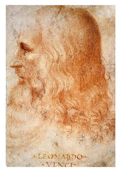
sea coasts, where all the shells were thrown up, broken.”Fig.1: Leonardo da Vinci at about age 60, drawn by his assistant Franceso Metzi in 1513.
Leonardo also specifically rejected the then popular view that shells had been left by the Biblical flood. "If the shells had been carried by the muddy deluge they would have been mixed up, and separated from each other amidst the mud, and not in regular steps and layers -- as we see them now in our time”. …”If the Deluge had carried the shells for distances of three and four hundred miles from the sea it would have carried them mixed with various other natural objects all heaped up together; but even at such distances from the sea we see the oysters all together and also the shellfish and the cuttlefish and all the other shells which congregate together, found all together dead; and the solitary shells are found apart from one another as we see them every day on the sea-shores.”
“And we find oysters together in very large families, among which some may be seen with their shells still joined together, indicating that they were left there by the sea and that they were still living when the strait of Gibraltar was cut through. In the mountains of Parma and Piacenza multitudes of shells and corals with holes may be seen still sticking to the rocks.”
If, as others then also believed, the shell forms had grown naturally in the rocks, Leonard wrote that "such an opinion cannot exist in a brain of much reason; because here are the years of their growth, numbered on their shells, and there are large and small ones to be seen which could not have grown without food, and could not have fed without motion -- and here they could not move."
Girolamo Fracastoro (1478 – 1553) was an Italian physician in Padua who was elected physician of the Council of Trent, and who was also a scholar in mathematics, geography and astronomy. In 1546 he proposed that epidemic diseases are caused by transferable tiny particles or "spores" that could transmit infection by direct or indirect contact or even without contact over long distances, an early theory of “germ” transmission of disease that had great influence for centuries. The transferable spore concept of Fracastoro is curiously analagous to the later molecular theory of the fossilization process as understood by Steno, involving the replacement of organic matter by stone.
With regard to fossils, Fracastoro also noticed shells and other marine fossils commonly found at upland locations such as around Padua, and also in Verona, where (as Lyell [1850] relates) many “curious petrifactions” had come to light during excavations in the city in 1517. Amid much speculation on the origin of the petrifactions, Fracastero stated that in his opinion the fossil shells were those of once living animals who had thrived in that place at a remote time.
Like Da Vinci, Fracastero used clear observations to refute the idea of the Biblical deluge as having moved the shells. The Flood, he reasoned, even if it had moved the shells, must have strewed them over the surface, not buried them at vast depths in the interior of mountains. Fracastero also ridiculed current theories such as that of "plastic force," which could fashion stones into organic forms.
While original thinkers like Shen Kuo, da Vinci, and Fracastero may not have been influenced by dogmatic religious beliefs, many others have been. In Europe, until the end of the 18th century, belief in the Biblical Deluge had notable influence on the interpretation of not only displaced seashells but also large vertebrate fossils. In southern England, for example, the cast of a small Jurassic dinosaur was found in a quarry in about 1700. It was locally interpreted as a person killed in the Flood, and the slab of rock was exhibited in the local church, where Robert Stukely found it in 1709 and identified it as a fossil reptile, later identified as an Iguanodon. In a similar but more famous case, a large fossil vertebrate found in Oeningen, Switzerland in 1725 was interpreted by a local naturalist named Scheuchzer to be a human witness of the Flood. This specimen, published by Scheuchzer in 1731, was proudly exhibited in Oeningen as "antediluvian man", until the French anatomist Georges Cuvier went there in 1805 and demonstrated that it was really a giant salamander.
Accuracy of observation on both living things and fossils.
Recording life and living things has been interesting to humans for a long time. The earliest records of life by humans are paintings and sculptures of animals by peoples in the latest phase of the Paleolithic or Old Stone Age
 period, many showing great naturalistic skill. These renderings, which
date from about 25,000-10,000 years ago, have been found in the Old
World from France (fig.2) to Russia, and in the New World in parts of
Brazil. These images are notable for their descriptive focus, in that
they record now extinct animals with careful attention to natural
details, so that many of the species, even extinct ones known otherwise
only from the fossil record, can be identified.
period, many showing great naturalistic skill. These renderings, which
date from about 25,000-10,000 years ago, have been found in the Old
World from France (fig.2) to Russia, and in the New World in parts of
Brazil. These images are notable for their descriptive focus, in that
they record now extinct animals with careful attention to natural
details, so that many of the species, even extinct ones known otherwise
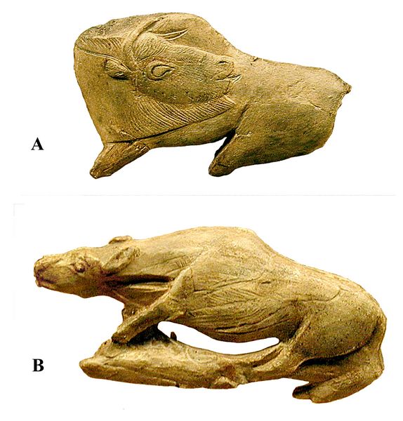
only from the fossil record, can be identified. Fig.2: Ivory carvings of animals by Magdalenian artists in France (15,000-12,000 years ago): A. auroch or wild ox; B. unidentified, possibly a lion (Musee National Prehistorique du France, Saint-Germain-en-Laye; photos: Athena Review).
Thereafter, during the Neolithic era, numerous plants and animals were domesticated, which clearly implies an ongoing, detailed knowledge of these species by many individuals around the world. Eventually, this knowledge was recorded for posterity. Writing, which began in Mesopotamia and Egypt by about 3500 BC, would become essential to the recording and spread of such knowledge about animals and plants.
Eventually, printing in both the Far East and Europe created wide access to descriptive sciences including natural history, and greatly facilitated comparative research. With printing, written descriptions could be accompanied by graphic illustrations for subjects ranging from medicine and human anatomy to herbals. The woodcuts or etchings which illustrated such texts during the early period of European printing in the Renaissance (about 1450-1600) are of variable quality, and sometimes subject to fantasy. They range from crude woodcuts in herbals and natural histories, often not detailed enough to serve for species distinctions, to highly skilled drawings by the likes of Albrecht Durer, Leonardo da Vinci, and Jan Stephen van Calcar, a student of Titian’s who illustrated Galen’s Anatomy.
16th century Natural Historians.
A precondition for any accurate rendering of the more fragmentary remains of fossils was a comparative data base of increasingly faithful representations of living organisms. The start of this coincided with the introduction of printing just after 1450. Book publication enabled the illustrated descriptions in one work to be readily compared with others, leading to a number of 16th century publications on natural history. Subjects of 16th century natural history were frequently botanical. Among the most accurate representations were in a 1520 herbal by the Italian Pacino Zenobia, who utilized plant pressing methods to get accurate drawings.
Specialized 16th century works on fish and other animals included the work of Pierre Belon (1517–1564), a French doctor and naturalist also known by the Latinized name Petrus Bellonius Cenomanus. Belon was a physician in Paris who travelled extensively and wrote detailed accounts of his journeys. He also wrote several original works on the comparative anatomy of fish and birds, including Histoire naturelle des estranges poissons (1551), De aquatilibus (1553), and L'Histoire de la nature des oyseaux (1555). One of his best known comparisons was between the skeleton of a bird, and that of a human (Gudger 1934).
Another French naturalist of the same time was Guillame Rondalet (1507-1566), a professor of medicine at
 the university of Montpellier. Rondalet’s major work was an illustrated
treatise on fish and other aquatic animals, Libri de piscibus marinis
in quibus verae piscium effigies expressae sunt. The work, published in
1556, covered not only fish (fig.3), but also marine mammals such as
seals and whales, and crustaceans and other invertebrates. Rondalet
dissected and illustrated numerous creatures, with his anatomical
drawing of a sea urchin is the earliest extant depiction of an
invertebrate. As a comparative anatomist, Rondalet found and reported
basic similarities between dolphins, pigs and humans (Gudger 1934).
the university of Montpellier. Rondalet’s major work was an illustrated
treatise on fish and other aquatic animals, Libri de piscibus marinis
in quibus verae piscium effigies expressae sunt. The work, published in
1556, covered not only fish (fig.3), but also marine mammals such as
seals and whales, and crustaceans and other invertebrates. Rondalet
dissected and illustrated numerous creatures, with his anatomical
drawing of a sea urchin is the earliest extant depiction of an
invertebrate. As a comparative anatomist, Rondalet found and reported
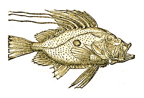
basic similarities between dolphins, pigs and humans (Gudger 1934).Fig.3: John Dory fish (Zeus faber, order Zeiformes), illustrated by Guillame Rondalet in his 1556 book Libri de piscibus marinis.
Contemporary with both Belon and Rondalet, and sometimes considered superior in his drawings of marine life, was Hippolyto Salviani (1514-1572), a professor of medicine at Rome. In his book Aquatilium animalium Historiae liber primus, cum Eorundem Formis, Aere Excusis (1554-1558) he portrayed fish mostly obtained in Roman fish markets (Gudger 1934). His approximately 100 drawings include detailed renderings of fish ranging from sharks and rays to sea scorpions (fig.4), and cephalopod species including squids and octopi. Most are from Italy, with a few examples from the Illyrian coast, along with several Mediterranean mollusks.
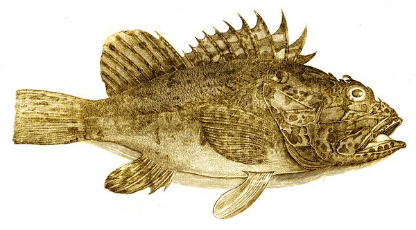 Fig.4: Sea Scorpion (order Scorpaeniformes) drawn by Hippolyto Salviani, from his book Aquatilium animalium Historiae (1558).
Such mid-to-late 16th century works on fish and other marine creatures by naturalists such as Belon, Rondalet, and Salviani reached relatively high levels of descriptive accuracy in both written and graphical formats. This was linked both to Renaissance artistic influences and to an increasingly naturalistic trend of detailed, analytic observations of many subjects.
Concurrently, in the same period there occurred the earliest known published drawing of a fossil in Europe. Although the writer did not realize it was a fossil, one of his readers did, which represented a kind of conceptual breakthough for academic geologists of that period. The fossil was a petrified mollusk shown in a 1557 book by Christoph Encellius (1517-1586), a German priest and highly regarded geologist. Encellius, in his book De Re Metallaci (On Metals), included sketches of two petrified objects resembling mollusks (fig.5), which were grouped by the author with gemstones and other minerals, as natural occurrences. This accorded with the then widespread belief that fossils were natural forms, which coincidentally resembled the forms of living creatures.
The following year, however, when the Swiss physician and naturalist Conrad Gessner, in Book 4 of his Historiae animalium (1558), compared the form illustrated by Encellius with an extant species of striated mollusk described by Guillame Rondelet in 1554, he arrived at a different conclusion. Gessner (1516-1565) is best known for his systematic and encyclopedic writings on animals and plants. His four volume Historiae animalium
 (1551-1558) covered quadrupeds, birds, fish, and snakes, with a final
volume “On Fossil Objects” (1565) describing fossils and minerals.
Comprising some 1297 pages and illustrated by over 900 woodcuts,
Gessner's work represents an important step towards modern zoology,
substantially aided by its level of scholarship. In Book 4, for
example, devoted to fish and other aquatic vertebrates, Gessner
incorporated the work of Belon, Rondalet, and all other known writers
on aquatic fauna, and labelled each form with the name of its
original describer (Gudger 1934).
(1551-1558) covered quadrupeds, birds, fish, and snakes, with a final
volume “On Fossil Objects” (1565) describing fossils and minerals.
Comprising some 1297 pages and illustrated by over 900 woodcuts,
Gessner's work represents an important step towards modern zoology,
substantially aided by its level of scholarship. In Book 4, for
example, devoted to fish and other aquatic vertebrates, Gessner
incorporated the work of Belon, Rondalet, and all other known writers
on aquatic fauna, and labelled each form with the name of its
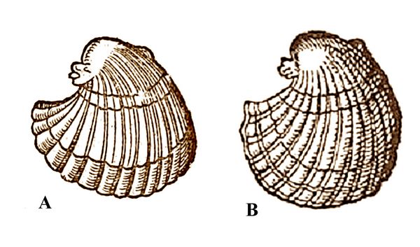
original describer (Gudger 1934). Fig.5: A) Drawing of petrified scallop-like shell by Encellius (1557), compared to B) a copy of the same drawing by Gessner (1558).
Gessner, when comparing the striated mollusks presented in the two separate works ofy Rondalet (1554) and Encellius (1557), recognized the petrified object in the Encellius figure as a fossil, as we understand the term (fig.5). In spite of the prevailing tendency, noted above, to interpret fossils as natural inclusions like minerals, Gessner was able to reach the alternative conclusion that some petrified forms, at least, were once living things.
A few years later, in his 1565 book “On Fossil Objects”, Gessner compared the closely similar forms of two echinoderms, a modern sea urchin and a lithified heart urchin, and concluded that the latter must have been a living form, and thus was an echinoderm fossil. Gessner, however, classed most fossils to be geological forms only
 resembling
living things. Two other lithified sea urchins which Gessner examined
in the same publication, which appeared rounder than the modern urchin,
and also differed in various other details, were judged to be natural
inclusions without an organic origin, which coincidentally resemble sea
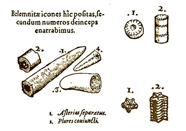
urchins. In a similar example (fig.6), Gessner considered the
tubular- shaped guards of belemnites and stacked peduncles of crinoids
to be minerals.
resembling
living things. Two other lithified sea urchins which Gessner examined
in the same publication, which appeared rounder than the modern urchin,
and also differed in various other details, were judged to be natural
inclusions without an organic origin, which coincidentally resemble sea
urchins. In a similar example (fig.6), Gessner considered the
tubular- shaped guards of belemnites and stacked peduncles of crinoids
to be minerals.Fig.6: Gessner's drawing of belemnite guards at left, which he called "arrow-shaped forms", and crinoid penducles at right (top: Encrinus liliformis; bottom: unidentified, star-shaped species), which Gessner considered as natural crystal forms (Gessner 1565).
Gessner’s mixed views underscore the historical point that, even for the most skilled naturalists of that era, familiar with details of many live animals and plants, there seemed usually no compelling reason to think their resemblance to petrified objects was any more than a mere coincidence. The fact that even a trained observer of nature like Conrad Gessner could not always be sure of the similarities between fossil and present-day sea urchins underscores one of the inherent problems of fossil identification. Except in the case of the most obvious fossils, such as shark's teeth, or the heart urchin illustrated by Gessner, or the very similar mollusks illustated by Rondalet (extant) and Encellius (fossil), the resemblance of often fragmentary or crushed fossils to living organisms is not often clear.

In terms of the physical materials of fossils, the necessary criteria for identifying fossils as opposed to inorganic inclusions such as rock crystals had not yet been established at the time of Gessner and his contemporaries, and would not be for a considerable period. Limitations here included a lack of previous references, published or in collections, allowing such comparisons; a lack of knowledge of how fossils were physically formed; and, given a lack of systematic geology, a grossly underestimated age of the earth, limiting the ability to imagine sufficient time depth involving former organisms turning to stone; or different terrains in past ages. .
Fig.7: Durer's 1515 drawing of a rhinocerous (Gessner 1558)
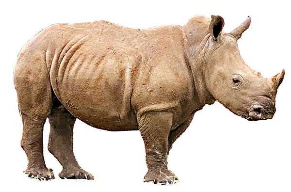
Gessner’s work, while fully illustrated, included drawings of mixed accuracy and level of detail. These ranged from relatively crude woodcut drawings to some elaborate renderings, such as Albrecht Durer’s 1515 drawing of a rhinocerous (fig 7). While aptly catching the pose of a white rhinocerous (fig.8), Durer's rendering is otherwise not as naturalistic as some of his other works, seeming to be almost a caricature of an animal with armor plating.
Fig.8: Photo of a modern white rhinocerous.
To resolve accuracy, both live and fossil specimens had to be drawn and understood in full detail, and students of biology and anatomy had to have acccess to the drawings. Printing solved the access part of the problem, but the discipline of comparative anatomy, essential to paleontology, took time to develop. Renaissance artists such as da Vinci provided the leading edge in human anatomy, and related comparative anatomy, but had to obtain corpses for study independently, and often illegally. Accurate formal training in human anatomy was only beginning in the 16th century, with the work of Vesalius and other anatomists to correct the methods of Galen; i.e., to separate human anatomy from that of the apes and pigs which the ancient Roman anatomists had used as proxies for human skeletons (see below).
The approach to accuracy in biological graphics (prior to photography) proceeded at different rates for different subjects. We have already seen that descriptive drawings of marine animals had reached a fairly high level by the mid to late 16th century, and this continued though the 17th and 18th centuries. Much the same can be said for botanical illustration. As discussed below, the introduction of the microscope in the early 17th century almost immediately led to a revolution in the description of small organisms, with many drawings of impressive accuracy dating from 17th and 18th century microscopic examinations.
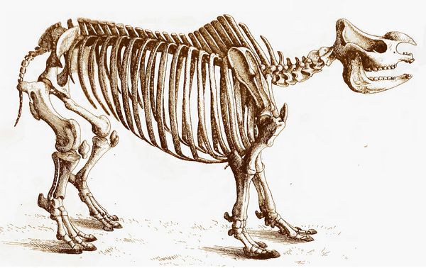
Fig.9: Cuvier's drawing of a rhinocerous skeleton (Cuvier 1812, vol.3)
For comparative anatomy of large land vertebrates, however, particularly of fossil land vertebrates (once they were identified as such), the pace lagged somewhat. With the exception of human anatomy, both the description of anatomical details and the drawings to illustrate them would not reach needed levels of accuracy until the late 18th century, with Stubbs and others in animal anatomy, and Cuvier and his contemporaries in the comparison of extant and fossil animals (fig.9).
Recording the human animal.
Perhaps the single most impressive set of drawings on human anatomy are contained in hundreds of pages of notes by Leonardo Da Vinci, drawn in between about 1489 and 1510, which remained unpublished upon his death in 1515, and unknown until the 19th century. A relatively complete collection of Leonardo's anatomy drawings were published in the 1950s. Most of da Vinci's illustrated notebooks, including the anatomy drawings, are owned by the Royal Windsor Collection at Buckingham Palace, where they were recently exhibited in 2012.
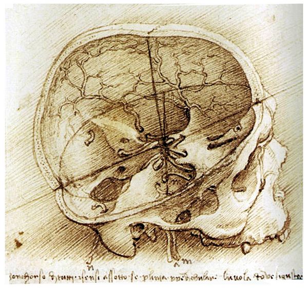 Fig.10: Drawing of a human skull
by da Vinci, ca.1490, showing the interior basicranial area with a
number of small openings (foramen) for nerves and blood vessels (image
reversed, to show labels and notes).
Fig.10: Drawing of a human skull
by da Vinci, ca.1490, showing the interior basicranial area with a
number of small openings (foramen) for nerves and blood vessels (image
reversed, to show labels and notes).Da Vinci is said to have dissected as many as 30 cadavers obtained from doctors and anatomy students. His accuracy of portraying detailed human skeletal and muscular features is comparable to, and in some cases considered superior to, that of Vesalius, whose drawings were used for centuries as a standard anatomy textbook (cf. Royal Library exhibit catalog, 2012). Da Vinci’s written observations on anatomy, placed among the drawings in his usual mirror script (fig.10), as were his views (described above) on geological formations and fossils, provide a classic case of simple but accurate observations serving as analytic steps to fuller understanding, limited by few preconceptions.
Andreas Vesalius (1514-1564), the latinized name of the Flemish Andries van Wesel, taught as a professor of anatomy at Padua, lecturing also at Bologna and Pisa. He replaced earler methods of relying on the classic textbooks of the ancient Roman anatomist Claudius Galen (AD 129-199) with dissection as the primary teaching tool. As in the case of Da Vinci, Nicholas Steno, and others who today would be regarded as scientists, direct observation was considered by Vesalius as the only reliable resource, representing a major break with medieval
 practices
of relying on prior authorities. He made many detailed anatomical
renderings and diagrams (fig.11) which he gathered into six tables of
anatomy for his students. After learning his work was widely copied, he
first published the complete set of drawings in 1538 under the title
Tabulae Anatomicae Sex.
practices
of relying on prior authorities. He made many detailed anatomical
renderings and diagrams (fig.11) which he gathered into six tables of
anatomy for his students. After learning his work was widely copied, he
first published the complete set of drawings in 1538 under the title
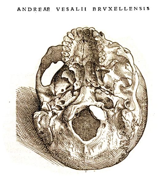
Tabulae Anatomicae Sex.Fig.11: Drawing of a human skull by Vesalius, ca.1538-40, showing the foramen magnum at the exterior base, where the spinal cord enters.
In 1539 Vesalius was able to obtain corpses of executed criminals for dissection. The same year he published an updated version of Galen's anatomical handbook, Institutiones Anatomicae. In 1541 Vesalius discovered, through manuscripts found in Bologna, that Galen’s anatomical research had been based on dissections of apes and pigs, rather than humans. Galen, originally from Pergamon in Turkey, but who had moved to Rome to be the physician of the emperors Commodus and Septimius Severus, had regarded apes as anatomically similar to humans. Since human dissection was banned in Rome, Galen used barbary apes as subjects. This led to further corrections by Vesalius of Galen’s Opera omnia.
Vesalius published his own seven volume text on human anatomy in 1543-4 under the title De humani corporis fabrica (On the fabric of the human body), with highly detailed illustrations. Many of these are thought to have been drawn by one of Titian's pupils, Jan Stephen van Calcar, who was present at dissections. Vesalius also published an abridged edition for students, Andrea Vesalii suorum de humani corporis fabrica librorum epitome. His observations showed various innaccuracies in Galen on human organs and skeletal anatomy. Among these were the functions and structure of the heart. Vesalius was the first to observe that the heart had four chambers, the liver two lobes, and that the blood vessels originated in the heart, not the liver. Vesalius also showed that the human lower jaw or mandible was a single bone (the dentary), not two as Galen had assumed from animal dissection.
The microscopic eye.
As students of ancient life soon discover, many plant and animal fossils are quite small, measuring only a few millimeters across (i.e., about 1/10 of an inch)—or less. Some of the most common organisms discovered are miniscule crustaceans called ostracods, which have flourished from the Ordovician era through today- for about 500 million years, and counting. These tiny creatures, contained within a bean-shaped shell usually less than 1 mm in length (fig.12), are much like miniature shrimp, with head, thorax, and multiple legs dangling below. They are
 found by the millions in tropical, coral reef environments today. In
fossil form their shells are also found in multitudes, spread out
across limestone formations which are actually sea floor remains of a
certain moment in the ancient past. Since ostracods are so
abundant, with a large nunber of species that changed over time, they
are highly useful index fossils to help date a given strata.
found by the millions in tropical, coral reef environments today. In
fossil form their shells are also found in multitudes, spread out
across limestone formations which are actually sea floor remains of a
certain moment in the ancient past. Since ostracods are so
abundant, with a large nunber of species that changed over time, they
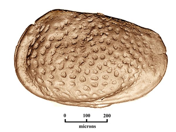
are highly useful index fossils to help date a given strata.Fig.12: The Late Jurassic ostracod Mandelstamia malacata, 88 microns long, is named for its spotted or pitted shell surface. M. malacata is a marker for the Kimmeridgean stage, 157-151 mya (photo after Natural History Museum (UK) cat. Hull-631).
Another small fossil type widely used in biostratigraphy today are conodonts (“cone-teeth”). They are found in marine deposits, commonly associated with graptolites, radiolarians, fish remains, brachiopods, cephalopods, trilobites, and ostracods. Conodonts for a long time were a puzzle to their discoverers.The name "conodont" was coined by the Latvian biologist C.H. Pander while working on Silurian fish fossils from Eastern Europe. Other conodont teeth were found on the ocean floor by the late 19th century British exploratory ship H.M.S. Challenger. Numerous species in North America were described by Ulrich and Bassler (1926), who were the first to recognise their biostratigraphic usefulness. In 1934 Schmidt and Scott discovered groups of individual elements preserved together in the same geological strata, suggesting they originally occurred in pairs, and could be mouth parts.
Finally, in the 1980s, conodont teeth were found together with bodily remains in a layer of fine grained Carboniferous shale in Scotland. The conodonts were revealed as miniature eel-like chordates, mostly about 3 cm long, although one genus from South Africa, Promissum, grew up to 16 inches long (fig.13C; Gabbott et al. 1995). Conodonts lacked a proper jaw, yet had an assemblage of tooth like elements inside their mouths for either filter feeding or catching food items (fig.13A,B). Because they had a notochord and block-like arrangement of muscles (called myomeres), conodonts are grouped with vertebrates, although the relation with other taxa is not yet well understood.
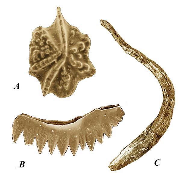
Fig.13: A, B: Condont teeth elements from the Early Silurian, Landovery stage. A. Gamachignathus macroexcavatus. Xiushan Formation, Guizhou Province, China. B, Apsidognathus tuberculatus, Wych Formation, Malvern Hills, UK. C: Promissum, Late Ordovician, Soom Shale, South Africa.
From the 1960s onwards, conodonts have developed into one of the most important biostratigraphic tools available in Palaeozoic and Triassic rocks. The very earliest conodonts are known from rocks of probable Precambrian age in Siberia. They appear more commonly in Cambrian deposits, with increasing diversity first in the Ordovician, and later, during the Devonian. The conodont-bearing organism survived the Permo-Triassic boundary extinctions but became extinct during the late Triassic. The extinction of conodonts coincides with the diversification of dinoflagellates and first appearance of calcareous nannofosils. Conodont tooth remains must be retrieved by fine sifting of limestone and placed on microscopic slides for viewing. Primarily on the basis of tooth morphology studied over the past 150 years, they have been split into 47 families and 180 genera. While major reclassification is underway now that they have been identified as chordates, conodonts remain the most commonly used index fossil to identify the date ranges of geological strata.
Invention of the microscope.
Fossilized or live organisms such as ostracods, conodonts, and many others are impossible to see clearly with the eye alone, let alone observe their considerable detail in order to distinguish their species. Before such species could even be recognized, let alone described, a major innovation was required – the invention of the microscope. This, coupled with further development of very accurate drawing techniques, occurred in Holland at the end of the 16th, and beginning of the 17th century, based on advances in lens making.
Magnification can be achieved by reflecting an image through a convex, or rounded lens, such as a transparent crystal. Such a crystal can also focus heat and use the sun's rays to set fire to tinder. The principle of magnifying glasses or lens (so-called because shaped like a lentil seed) had been understood since Roman times, being mentioned by Seneca and Pliny the Elder in the 1st century AD. From such lens, eye glasses were made by the late 13th century.
Leonardo da Vinci, as revealed in his notebooks, conducted experiments with light travelling though a pinpoint hole or camera obscura, casting a reversed image on a wall behind. Passing an image through drops of water could magnify an image. From this Leonardo made (effectively, correct) inferences about light being transmitted in parallel waves. The images of what the eye sees are composed of (or, as Leonardo phrased it, are carried on) such parallel waves of light. If the parallels are distorted, the refocused image is enl
 arged
(or reduced, as when looking though the outer end of a telescope). The
eye itself contains such a convex lens for focusing our vision.
arged
(or reduced, as when looking though the outer end of a telescope). The
eye itself contains such a convex lens for focusing our vision.Among the first successful magnification lens were those created in about 1590 by Zaccharias Janssen and his son Hans Janssen, who made lens for eye glasses. They found that objects could be greatly magnified with two lenses placed in a tube, the basic principle of both the compound microscope and of the telescope. Hans Jensen later supplied lens to the Italian scientist Galileo Galilei, who used them to construct both microscopes and telescopes, his use of the latter proving a milestone in astronomy. In 1609, Galileo, based on the Jensens’ earlier experiments, studied the principles of lenses for application in astronomy. He built a more effective telescope capable of resolving detail on the surface of the Moon (which he drew), and in 1621 was the first to see the four largest satellites of the planet Jupiter, now called the Galilean satellites.
Fig.14: Drawing of the spinal cord of a cow by Antonie von Leeuwenhoek (1717). At top (1) is a lengthwise section of the cow`s spinal cord showing a nerve cord branching off. Below (2) is a cross section of the spinal cord nerve, showing five bundled nerves and surrounding fat.
Meanwhile, back in Holland, by the mid-to-late 17th century magnifying lens had been much improved by Antonie von Leeuwenhoek, who began as a draper's assistant and went on to revolutionize the science of biology through use of the microscope. Von Leeuwenhoek had noticed that drapers examined cloth fibers through
 magnifying lens. He copied
and improved these, making small, ball-like glass lenses that could
magnify up to 150 or 200 times. Von Leeuwenhoek
designed apparatus (single-lens microscopes) which enhanced the viewing
light and allowed him to observe and draw previously unseen subjects
thus magnified, such as the inner components of seeds (i.e., tiny
incipient plant parts), and the different layers of wood stems. During
the 1670s, he was the first to see and describe single-celled organisms
(protists), bacteria, sperm, cell vacuoles, and the banded structure of
muscle tissue (fig.14). He described his findings in a series of over
100 letters to the Royal Society in England, continuing to expand his
observations until age 90.
magnifying lens. He copied
and improved these, making small, ball-like glass lenses that could
magnify up to 150 or 200 times. Von Leeuwenhoek
designed apparatus (single-lens microscopes) which enhanced the viewing
light and allowed him to observe and draw previously unseen subjects
thus magnified, such as the inner components of seeds (i.e., tiny
incipient plant parts), and the different layers of wood stems. During
the 1670s, he was the first to see and describe single-celled organisms
(protists), bacteria, sperm, cell vacuoles, and the banded structure of
muscle tissue (fig.14). He described his findings in a series of over
100 letters to the Royal Society in England, continuing to expand his
observations until age 90.Fig.15: Drawing of a flea by Robert Hooke (1665).
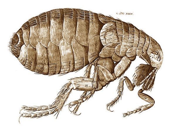
Von Leeuwenhoek’s early successes were followed by those of another gifted observer with good drawing skills, Robert Hooke of England, who in 1665 published the first book on the subject, Micrografica. Hooke’s graphic recordings of magnified wood cells, enlarged insects, and compound insect eyes, revealing completely new detail, reached a wide public (fig.15). Adoption of the microscope to biology ennabled vastly improved species descriptions. This paved the way for the microscopic study of small fossils, and enabled the first accurate definitions of Paleozoic organisms during the late 18th and early 19th centuries.
References:
Agricola, G. 1546 De veteribus et novis metallis (de natura Fossilium).
Aristotle Meteorologia
Avicenna 1027 Book of Healing
Belon, P. 1551. Histoire naturelle des estranges poissons.
Belon, P. 1553. De aquatilibus.
Belon, P. 1555. L'Histoire de la nature des oyseaux.
Da Vinci, L. Notebooks.
Da Vinci, L. Antomical Drawings. Royal Library, 2012.
Du Yu
Encellius, C. 1557. De Re Metallaci.
Fracastero
Gabbott, S.E.; R. J. Aldridge, J. N. Theron 1995. "A giant conodont with preserved muscle tissue from the Upper Ordovician of South Africa". Nature 374, pp. 800–803.
Gessner, C. 1552-1558 Historiae animalium (History of Animals) 4 vols.
Gessner, C. 1565 On Fossil Objects.
Gudger, E.W. 1934 "The Five Great Naturalists of the Sixteenth Century: Belon, Rondelet, Salviani, Gesner and Aldrovandi: A Chapter in the History of Ichthyology." Isis 22,(1), pp. 21-40.
Hooke, R. 1665. Micrografica.
Lyell, C. 1833, 1850 Elements of Geology.
Rondalet, G. 1556 Libri de piscibus marinis in quibus verae piscium effigies expressae sunt.
Royal Library 2012. "The Anatomical Drawings of Leonardo da Vinci." Catalog for exhibition of Windsor Collection.
Salviani, H. 1554-1558. Aquatilium animalium Historiae liber primus, cum Eorundem Formis, Aere Excusis.
Scheuchzer 1731
Shen Kuo
Shen Kuo Menxi Bitan (“Dream Pool”).
Steno, N. 1667. Nicolai Stenonis Elementorum Myologiae Specimen, seu Musculi Descriptio Geometrica, cui accedunt canis carchariae dissectum caput et dissectus piscis ex canum genere... Florentiae : ex typ. sub signo Stellae, via Bibliothèque interuniversitaire de médecine, Paris.
von Leeuwenhoek, Antonie, Letters . Published by the Royal Society.
Vesalius, A. 1538. Tabulae Anatomicae Sex.
Vesalius, A. 1543-4 De humani corporis fabrica (On the fabric of the human body).
Xenophanes
Zenobia, P. 1520.
Glossary